
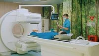
The silent progression of cancer to the skeletal system represents one of the most critical challenges in oncological care, particularly for patients diagnosed with breast or prostate malignancies. While bone metastasis often remains asymptomatic in its nascent stages, its impact on a patient’s long-term prognosis and quality of life is profound. To address the complexities of identifying these lesions, medical institutions are increasingly turning to advanced molecular imaging techniques, specifically Single-Photon Emission Computed Tomography combined with Computed Tomography (SPECT-CT), to provide a precise, high-resolution roadmap for diagnosis and treatment planning.
The clinical necessity for such advanced imaging stems from the nature of bone metastasis. When cancer cells migrate from primary sites to the bone, they initiate a cycle of bone destruction and disorganized bone formation. This process can lead to severe skeletal-related events, including debilitating pain, pathological fractures, and, in cases involving the spinal column, spinal cord compression. Detecting these changes before they manifest as clinical crises is the primary objective of modern nuclear medicine.
The Mechanism of SPECT-CT Imaging
SPECT-CT is a hybrid imaging technology that integrates functional and anatomical data. While a standard bone scan provides information about metabolic activity—highlighting areas where bone turnover is elevated—it often lacks the anatomical specificity required to distinguish between cancerous lesions and benign bone changes, such as degenerative disease or previous injuries.
By superimposing the functional imaging from the SPECT scan onto the precise anatomical imagery of a CT scan, physicians obtain a three-dimensional, fused view of the skeletal structure. This allows clinicians to pinpoint the exact location of abnormal activity with millimeter-level accuracy. This dual-modality approach significantly reduces the "gray area" of diagnostic ambiguity, providing a clear distinction between metastatic activity and other skeletal pathologies.
Clinical Perspective: Evaluating Bone Integrity
Dr. Yustia Tuti, Sp.KN-TM, Subsp.Onk (K), a specialist at Mayapada Hospital Jakarta Selatan, emphasizes that the utility of SPECT-CT extends far beyond simple detection. According to Dr. Yustia, the clinical priority is to develop a comprehensive understanding of the patient’s skeletal health to preempt potential complications.
"Oncological management requires a granular view of the patient’s status," Dr. Yustia explains. "When we identify bone metastasis, we are looking for more than just the presence of a lesion. We evaluate the extent of the spread, the specific location, and the mechanical integrity of the affected bone. This information is the bedrock upon which we build a targeted therapeutic strategy."
The implications of this data are significant. By understanding the extent of skeletal involvement, the multidisciplinary medical team can intervene before a pathological fracture occurs, which is a major factor in maintaining a patient’s mobility and independence. For patients with high-risk disease, early detection via SPECT-CT can be the difference between palliative intervention and proactive, life-extending management.
Distinguishing Pathology from Benign Findings
One of the most persistent hurdles in oncological imaging is the interpretation of "hot spots" on bone scans. These indicators, while often related to metastasis, can also be triggered by non-malignant conditions, including osteoarthritis, fractures, or inflammatory processes.
Dr. Andreas Lim, Sp.KN-TM, Subsp. Onk. (K), FANMB, also of Mayapada Hospital Jakarta Selatan, highlights the diagnostic precision afforded by the hybrid nature of the SPECT-CT system. "The primary strength of the SPECT-CT scan lies in its ability to clarify findings that would otherwise be ambiguous on conventional imaging," states Dr. Lim. "By correlating the increased metabolic uptake seen in the SPECT phase with the anatomical details from the CT phase, we can confidently categorize a lesion as metastatic or benign. This level of clarity prevents unnecessary anxiety for the patient and ensures that the subsequent treatment path is both accurate and necessary."
The Evolution of Cancer Care in Indonesia
The integration of advanced nuclear medicine into mainstream oncology reflects a broader shift in the Indonesian healthcare landscape. Historically, patients requiring advanced molecular imaging often faced limited access or necessitated long-distance travel. The current deployment of SPECT-CT technology within facilities such as the Poli Kedokteran Nuklir at Mayapada Hospital Jakarta Selatan represents a strategic investment in the country’s diagnostic infrastructure.
This move toward localized, high-tech diagnostics aligns with the global trend of "Comprehensive Cancer Care." This model emphasizes that imaging should not exist in a vacuum but must be integrated into a larger ecosystem of care. In this model, the results from a SPECT-CT scan are presented to a "Tumor Board"—a multidisciplinary team of surgeons, medical oncologists, radiation oncologists, and nuclear medicine specialists—who collaborate to create a personalized treatment plan.
Broader Implications for Patient Outcomes
The implementation of SPECT-CT is not merely a technological upgrade; it is a clinical intervention that directly impacts survival rates and quality of life. The data derived from these scans influences key therapeutic decisions, such as the timing of bisphosphonate therapy, the necessity for prophylactic orthopedic surgery to stabilize vulnerable bones, or the adjustment of systemic chemotherapy regimens.
Moreover, the psychological impact of having a clear, evidence-based diagnostic path cannot be overstated. Patients navigating a cancer diagnosis often face a period of extreme uncertainty. Providing patients with a definitive assessment of their skeletal health, managed by a team that utilizes the most sophisticated diagnostic tools available, fosters a collaborative relationship between the patient and the medical institution.
Integrating Technology and Holistic Monitoring
Beyond the specialized care offered in the oncology department, the medical industry is increasingly integrating patient-facing technology to monitor general health. Digital health ecosystems, such as the MyCare application, exemplify this trend. By offering features that track physical indicators like heart rate, caloric expenditure, and BMI, these platforms encourage a level of patient engagement that is essential for overall wellness.
While these tools are distinct from the highly specialized domain of nuclear medicine, they represent the same core philosophy: the use of data to inform better health outcomes. For a cancer patient, maintaining a baseline of physical health is vital to ensuring they remain robust enough to undergo intensive treatments, including surgery, radiation, or chemotherapy.
Looking Ahead: The Future of Metastatic Management
The future of bone metastasis management lies in the continued refinement of molecular imaging and its integration into early-stage diagnostic workflows. As artificial intelligence begins to assist in the interpretation of complex medical imagery, the speed and accuracy of SPECT-CT diagnostics are expected to increase further.
For now, the priority remains the widespread adoption of these tools to ensure that patients in Indonesia have equitable access to state-of-the-art care. The collaboration between nuclear medicine specialists and the broader oncology team—supported by resources like the Patient Navigator program, which guides patients through the intricacies of their treatment journey—serves as a model for sustainable and patient-centered healthcare.
Patients and families seeking more information regarding these services or requiring guidance on navigating a cancer diagnosis are encouraged to utilize available support channels. Accessing specialized services through verified contact points, such as the Mayapada Hospital call center (150770) or their dedicated emergency services (150990), ensures that patients receive accurate, timely, and professional assistance tailored to their specific medical requirements.
Conclusion
The challenge of bone metastasis remains a significant hurdle in the fight against cancer. However, the advancement of SPECT-CT technology provides a critical tool for clinicians to "see" the unseen, enabling them to transition from reactive treatment to proactive, evidence-based management. By combining the high-resolution anatomical imaging of CT with the functional insights of nuclear medicine, hospitals are providing a new standard of care that prioritizes both the survival and the long-term well-being of the patient. Through continued investment in medical technology and a commitment to multidisciplinary collaboration, the oncology community remains better equipped than ever to face the complexities of metastatic disease.